Study shows signs of atrial fibrillation from eye scans four years before diagnosis
People with atrial fibrillation (AF) have distinctive biomarkers in their retinas, evident as early as four years before clinical presentation of the condition, confirms a new study published in PLOS Digital Health, led by Moorfields Eye Hospital NHS Foundation Trust and UCL Institute of Ophthalmology.
The research team analysed retinal imaging data from more than 90,000 people and discovered differences in the structure of the retina in people with AF, compared with those who do not have the condition.
AF, the most common heart rhythm disorder, affects more than 1.5 million people in the UK, and substantially raises the risk of stroke, heart failure and death. Because AF often occurs intermittently, it can be missed on a standard electrocardiogram (ECG). As a consequence, many cases remain undiagnosed until a serious or life-threatening complication occurs.
Moorfields patient John Brewer first discovered he had AF in the operating theatre, while undergoing a cataract operation under sedation, earlier this year. The surgical team noticed John was experiencing heart arrhythmia and paused the procedure to hook up an ECG, which indicated that John did have AF. Medication was then provided so the operation could be safely completed. John was given an immediate referral to see a cardiologist who has since confirmed the AF diagnosis.
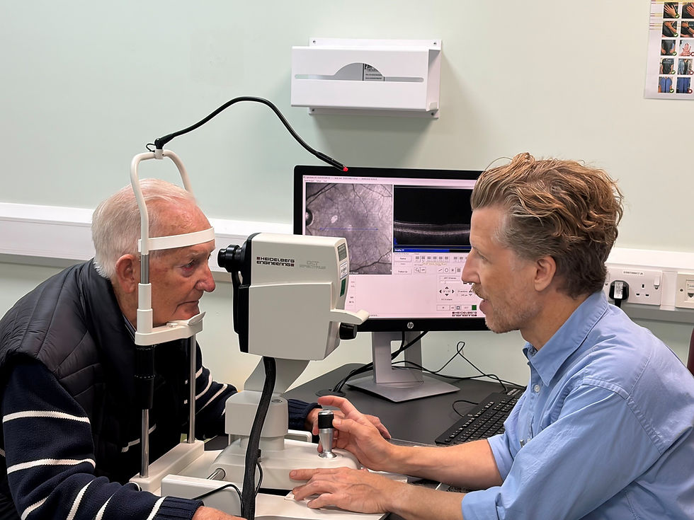
John said “It would have been so much easier if my AF had been detected earlier through a routine retinal scan at one of my regular appointments, rather than on the operating table, which was a very distressing experience for my wife and I.”
The Moorfields team set out to establish whether retinal imaging, already used routinely in community optometry practices and hospital eye clinics, could offer a non-invasive way to flag people with AF before they have a stroke or heart attack.
Researchers analysed optical coherence tomography (OCT) scans and colour retinal photographs from two datasets. In the AlzEye dataset, a Moorfields cohort with over 50,000 patients, 5,735 already had AF. In the UK Biobank cohort of around 40,000 participants, 526 had AF. Across both datasets, people with AF consistently had thinner macular ganglion cells and inner plexiform layer (mGCIPL), which is a retinal layer at the back of the eye made up of nerve cells that connect the eye to the brain. The thinner this particular layer was, the more likely the person was to be diagnosed with AF.

Notably, among patients who did not have AF when their retinal images were taken, those who went on to develop AF had thinner mGCIPL layers on average four years before presenting to hospital with the condition, even after accounting for age, sex, ethnicity, hypertension, diabetes, smoking and alcohol use. This indicates that retinal changes may be detectable before a clinical diagnosis of AF is made, at a stage when the condition has no symptoms.
The authors propose that the thinning they observed in people who developed or had AF could indicate mini strokes in the brain that typically occur as a result of AF before its diagnosis. The thinning could happen because blood flow to the retinal nerve cells is reduced by these strokes.
Senior author Siegfried Wagner said: “This large-scale analysis shows that retinal imaging can pick up on structural changes within the eye that are clearly linked to AF. For patients, the value of this kind of eye imaging is that it is easy to access, quick and doesn't involve any invasive procedures. Although not intended to replace ECG-based diagnosis, retinal imaging could act as a complementary tool, catching those most at risk of AF before it causes real harm.”
Lead author Josef Huemer said: “If confirmed in further research, our findings raise the possibility that routine eye examinations could, in future, help identify people who would benefit from closer cardiac assessment, potentially enabling earlier treatment with therapies known to reduce the risk of stroke.”
This study adds to growing evidence that high-resolution images of the eye can be used for early detection of systemic disease, a scientific field known as oculomics. The Moorfields team has been able to identify early signs and predict progression risk of eye conditions including AMD, glaucoma and diabetic retinopathy. The same team has also found markers for Parkinson’s disease, Alzheimer’s disease, schizophrenia, cardiovascular disease and stroke.
Oculomics has been made possible by the INSIGHT Eye and Oculomics Health Data Research Hub at Moorfields Eye Hospital NHS Foundation Trust and UCL Institute of Ophthalmology. INSIGHT is the world’s largest ophthalmic imaging bioresource, with over 30 million eye images linked to medical records.
For more information email enquiries@insight.hdrhub.org
Research collaborators
The research was supported by Moorfields Eye Charity, NIHR Biomedical Research Centre: Moorfields, NIHR Biomedical Research Centre: University College London Hospitals, NIHR Biomedical Research Centre: Great Ormond Street Hospital, NIHR Biomedical Research Centre: Barts, NIHR Biomedical Research Centre: Birmingham
About Moorfields Eye Hospital NHS Foundation Trust Moorfields Eye Hospital NHS Foundation Trust is one of the leading providers of eye health services in the UK and a world class centre of excellence for ophthalmic research and education. Our main focus is the treatment and care of NHS patients with a wide range of eye problems, from common complaints to rare conditions that require treatment not available elsewhere in the UK. Our unique patient case mix and the number of people we treat mean that our clinicians have expertise in discrete ophthalmic sub-specialties. We treat people in 20 locations in and around London, the south east and Bedford, enabling us to provide expert treatment closer to patients’ homes. We also operate commercial divisions that provide care to private patients in both London and the Middle East.
With our academic partners at the UCL Institute of Ophthalmology, Moorfields is recognised as a leading centre of excellence in eye and vision research. Together we form one of the largest ophthalmic research sites in the world, with the largest patient population in Europe or the USA. We publish more scientific papers than any other eye and vision research site and have an extensive joint research portfolio. For more information please visit https://www.moorfields.nhs.uk/
About UCL Institute of Ophthalmology
UCL Institute of Ophthalmology is one of a number of specialised research centres within UCL (University College London) and is, together with Moorfields Eye Hospital, one of the leading centres for eye research worldwide. The combination of the institute’s research with the resources of Moorfields Eye Hospital opens the way for advances at the forefront of vision research. Close collaboration with other academic partners and with industry extends its impact. The institute has been named as the best place to study ophthalmology in the 2017 Centre for World University Rankings (CWUR). For more information, please visit www.ucl.ac.uk
About the INSIGHT Eye and Oculomics Health Data Research Hub
INSIGHT is the world's largest ophthalmic imaging bioresource, with over 30 million eye images linked to clinical data. An NHS initiative founded in 2019 and led by Moorfields Eye Hospital, INSIGHT makes routinely collected patient eye data available for approved research that could lead to improvements in diagnosis, care and treatment of eye diseases, as well as systemic disease such as stroke and dementia. INSIGHT’s mission is to improve healthcare for the benefit of patients, the NHS and wider society by enabling safe and trusted research access to anonymised data for research and innovation. For more information, please visit https://www.insight.hdrhub.org/
About NIHR Biomedical Research Centre: Moorfields
NIHR Biomedical Research Centre: Moorfields is an internationally renowned partnership between Moorfields Eye Hospital NHS Foundation Trust and UCL Institute of Ophthalmology, ranked number one in the world for ophthalmology research. The BRC was first established in April 2007 and is now in the fourth five-year term to support research designed to take advances in basic medical research from the laboratory to the clinic. The centre’s broad spectrum of research into eye disease enables patients to benefit more quickly from world-class scientific breakthroughs, treatments and diagnostics, and has demonstrated a wider reach into other health conditions through digital technology and advanced therapies. For further information, please visit www.moorfieldsbrc.nihr.ac.uk/
About AlzEye
The first health data project of its kind, AlzEye links more than 6 million retinal images from over 350,000 people attending Moorfields Eye Hospital (1997 - 2018) with nationally held data (NHS Digital) on people who develop Alzheimer’s disease and other forms of dementia.
Led by Siegfried Wagner and Pearse Keane, AlzEye provides a unique opportunity to explore patterns of retinal change associated with dementia development across a diverse population of varying ethnicity and socioeconomic status.
In 2025, an update to AlzEye was agreed with NHS England, updating the Hospital Episode Statistics (HES) data to 2024 and including cause of death certificates.For more information visit https://www.insight.hdrhub.org/alzeye